Journals > > Topics > Medical Optics and Biotechnology
Medical Optics and Biotechnology|257 Article(s)
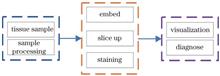
Breast Cancer Cell Recognition System Based on Microscopic Image
Guohua Liu, Keding Yan, Jing Xing, Guojun Ma, Jing Liu, and Yanli Chen
At present, it is highly subjective for pathologists to identify breast cancer cells in pathological cut images of breast cancer under microscope field of view by naked eyes. Therefore, we design a microscopic image based breast cancer cell recognition system, which includes microscopic image acquisition module and breast cancer cell recognition algorithm implementation module. Through USAF 1951 resolution test board, the microscopic image acquisition module of designed breast cancer recognition system is verified, and the final imaging resolution can reach 2.19 μm. In addition, the designed breast cancer cell recognition algorithm is verified by multiple sets of breast cancer pathological images, and the results show that the average accuracy of the designed breast cancer cell recognition system reaches 93.4%. At present, it is highly subjective for pathologists to identify breast cancer cells in pathological cut images of breast cancer under microscope field of view by naked eyes. Therefore, we design a microscopic image based breast cancer cell recognition system, which includes microscopic image acquisition module and breast cancer cell recognition algorithm implementation module. Through USAF 1951 resolution test board, the microscopic image acquisition module of designed breast cancer recognition system is verified, and the final imaging resolution can reach 2.19 μm. In addition, the designed breast cancer cell recognition algorithm is verified by multiple sets of breast cancer pathological images, and the results show that the average accuracy of the designed breast cancer cell recognition system reaches 93.4%.
Laser & Optoelectronics Progress
- Publication Date: Apr. 25, 2024
- Vol. 61, Issue 8, 0817001 (2024)

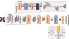
Research on Combining Self-Attention and Convolution for Chest X-Ray Disease Classification
Xin Guan, Jingjing Geng, and Qiang Li
Chest X-rays are used to diagnose a wide range of chest conditions. However, due to the complicated and diverse features of thoracic diseases, existing disease classification algorithms for chest radiographs have difficulty in learning the complex discriminating features of thoracic diseases and do not fully consider correlation information between different diseases. This study proposes a disease classification algorithm that combines self-attention and convolution to address these problems. This study employs omni-dimensional dynamic convolution to replace the standard convolution of the residual network to enhance the feature extraction capabilities of the network for multi-scale information. In addition, a self-attention module is introduced into the convolutional neural network to provide global receptive fields that capture correlations between multiple diseases. Finally, an efficient double path attention is proposed that allows the network to give greater attention to the focal area and automatic capturing of changes in lesion locations. The proposed model is evaluated on the ChestX-ray14 dataset. Experimental results show that the accuracy of the algorithm and the efficiency of diagnosis for the classification of 14 chest diseases is improved over those of the seven current state-of-the-art algorithms, with an average area under receiver operating characteristic curve (AUC) value of 0.839. Chest X-rays are used to diagnose a wide range of chest conditions. However, due to the complicated and diverse features of thoracic diseases, existing disease classification algorithms for chest radiographs have difficulty in learning the complex discriminating features of thoracic diseases and do not fully consider correlation information between different diseases. This study proposes a disease classification algorithm that combines self-attention and convolution to address these problems. This study employs omni-dimensional dynamic convolution to replace the standard convolution of the residual network to enhance the feature extraction capabilities of the network for multi-scale information. In addition, a self-attention module is introduced into the convolutional neural network to provide global receptive fields that capture correlations between multiple diseases. Finally, an efficient double path attention is proposed that allows the network to give greater attention to the focal area and automatic capturing of changes in lesion locations. The proposed model is evaluated on the ChestX-ray14 dataset. Experimental results show that the accuracy of the algorithm and the efficiency of diagnosis for the classification of 14 chest diseases is improved over those of the seven current state-of-the-art algorithms, with an average area under receiver operating characteristic curve (AUC) value of 0.839.
Laser & Optoelectronics Progress
- Publication Date: Feb. 25, 2024
- Vol. 61, Issue 4, 0417002 (2024)
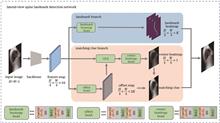
Lateral Spine Landmark Detection Based on Matching Clue Regression
Menghao Gao, Lijun Guo, Rong Zhang, Lixin Ni, Qiang Wang, and Xiuchao He
In lateral spine landmark detection, the previous heatmap regression methods have difficulty in distinguishing landmarks on different vertebrae due to the influence of organ occlusion and are prone to landmark and vertebrae matching errors. To solve this problem, we propose a new one-stage lateral spine landmark detection method, which simultaneously predicts the landmark heatmap and landmark matching clue (vertebra center heatmap and landmark offset), and uses the matching clue to match the landmarks with the corresponding vertebra. In order to improve the matching effect, we propose the geometry-aware feature aggregator module, which can extract the landmark features on the vertebra to enhance the feature representation of the vertebra center. We also use a weighted loss function to alleviate the imbalance of positive and negative samples in the landmark and the vertebra center heatmaps. Experimental results show that the average detection error of the proposed method is 8.84, which has 36% improvement in accuracy compared to the method with the second-highest performance. In lateral spine landmark detection, the previous heatmap regression methods have difficulty in distinguishing landmarks on different vertebrae due to the influence of organ occlusion and are prone to landmark and vertebrae matching errors. To solve this problem, we propose a new one-stage lateral spine landmark detection method, which simultaneously predicts the landmark heatmap and landmark matching clue (vertebra center heatmap and landmark offset), and uses the matching clue to match the landmarks with the corresponding vertebra. In order to improve the matching effect, we propose the geometry-aware feature aggregator module, which can extract the landmark features on the vertebra to enhance the feature representation of the vertebra center. We also use a weighted loss function to alleviate the imbalance of positive and negative samples in the landmark and the vertebra center heatmaps. Experimental results show that the average detection error of the proposed method is 8.84, which has 36% improvement in accuracy compared to the method with the second-highest performance.
Laser & Optoelectronics Progress
- Publication Date: Feb. 25, 2024
- Vol. 61, Issue 4, 0417001 (2024)
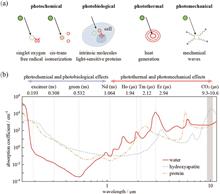
Research Progress and Perspective of Clinically Promising Flexible CO2 Laser Delivery Mediums (Invited)
Guangming Tao, Yuqi Zou, Chao Liu, and Zhihe Ren
Mid-infrared (MIR) lasers offer substantial benefits, including non-contact operation, high efficiency, and precision, making them widely utilized in clinical surgical procedures such as lesion tissue removal, tissue plasticity, and tumor interstitial photothermal therapy. Notably, carbon dioxide (CO2) lasers, among various MIR lasers, are extensively employed in skin, ear, nose, throat, and abdominal surgeries due to their exceptionally high ablation efficiency and precision. However, the lack of stable and high-performance small-scale, flexible laser energy-delivering mediums for CO2 lasers restricts their use in minimally invasive or noninvasive procedures, a capability present in mature silica fibers used in holmium, neodymium, and other near-infrared lasers for conducting minimally invasive interventional operations in natural cavities in vivo. Presently, CO2 laser procedures typically rely on energy-delivering mediums such as articulated arms and hollow waveguides but this considerably hampers the application of CO2 laser in minimally invasive surgeries. To enhance the role of CO2 lasers in clinical medicine, we review and summarize existing medical CO2 laser energy-delivering mediums, focusing on the advances in thermal-drawn multi-material fiber technology in CO2 laser surgery, and explore future development trends and applications of multifunctional flexible CO2 laser ablation robotic fibers. Mid-infrared (MIR) lasers offer substantial benefits, including non-contact operation, high efficiency, and precision, making them widely utilized in clinical surgical procedures such as lesion tissue removal, tissue plasticity, and tumor interstitial photothermal therapy. Notably, carbon dioxide (CO2) lasers, among various MIR lasers, are extensively employed in skin, ear, nose, throat, and abdominal surgeries due to their exceptionally high ablation efficiency and precision. However, the lack of stable and high-performance small-scale, flexible laser energy-delivering mediums for CO2 lasers restricts their use in minimally invasive or noninvasive procedures, a capability present in mature silica fibers used in holmium, neodymium, and other near-infrared lasers for conducting minimally invasive interventional operations in natural cavities in vivo. Presently, CO2 laser procedures typically rely on energy-delivering mediums such as articulated arms and hollow waveguides but this considerably hampers the application of CO2 laser in minimally invasive surgeries. To enhance the role of CO2 lasers in clinical medicine, we review and summarize existing medical CO2 laser energy-delivering mediums, focusing on the advances in thermal-drawn multi-material fiber technology in CO2 laser surgery, and explore future development trends and applications of multifunctional flexible CO2 laser ablation robotic fibers.
Laser & Optoelectronics Progress
- Publication Date: Jan. 10, 2024
- Vol. 61, Issue 1, 0117001 (2024)
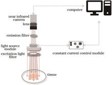
Rapid Parathyroid Recognition System Based on Near-Infrared Autofluorescence
Chunhui Yao, Yang Zhang, Bin Liu, Chijian Zhang, Jiayun Zheng, Xia Wang, Xu Kang, Quanfu Wang, Zhongsheng Li, Yong Liu, Meili Dong, and Yikun Wang
Based on near-infrared autofluorescence, a rapid identification system for parathyroid glands during surgery is designed, the system is of great value for rapid identification of parathyroid glands during operation. In this research, a ring-shaped adjustable excitation light source and a high-precision adjustable LED constant current source are designed. The near-infrared light source is used to excite tissue fluorescence, and the tissue autofluorescence information is collected by a high-sensitivity CMOS camera. The obtained fluorescence images are processed, accurately identifying parathyroid glands. Simulating tissue fluorescence through gradient concentration of indocyanine green (ICG) solution, the experimentally measured fluorescence intensity is positively correlated with the concentration of ICG, and both the signal-to-noise ratio and signal-to-background ratio meet the requirements for intraoperative discrimination, which verifie the sensitivity and accuracy of the proposed system for different fluorescence intensities. Using this system to test tissue phantoms, the fluorescent phantom can be clearly distinguished from the background. The parathyroid gland and surrounding tissues were tested, and the parathyroid gland is green and clearly distinguished from the surrounding tissues, which preliminarily verifies that the proposed system can be used for the identification and detection of parathyroid glands. Based on near-infrared autofluorescence, a rapid identification system for parathyroid glands during surgery is designed, the system is of great value for rapid identification of parathyroid glands during operation. In this research, a ring-shaped adjustable excitation light source and a high-precision adjustable LED constant current source are designed. The near-infrared light source is used to excite tissue fluorescence, and the tissue autofluorescence information is collected by a high-sensitivity CMOS camera. The obtained fluorescence images are processed, accurately identifying parathyroid glands. Simulating tissue fluorescence through gradient concentration of indocyanine green (ICG) solution, the experimentally measured fluorescence intensity is positively correlated with the concentration of ICG, and both the signal-to-noise ratio and signal-to-background ratio meet the requirements for intraoperative discrimination, which verifie the sensitivity and accuracy of the proposed system for different fluorescence intensities. Using this system to test tissue phantoms, the fluorescent phantom can be clearly distinguished from the background. The parathyroid gland and surrounding tissues were tested, and the parathyroid gland is green and clearly distinguished from the surrounding tissues, which preliminarily verifies that the proposed system can be used for the identification and detection of parathyroid glands.
Laser & Optoelectronics Progress
- Publication Date: Mar. 25, 2023
- Vol. 60, Issue 6, 0617003 (2023)

Improved YOLOv4 Model-Based Spinal Magnetic Resonance Imaging Image Detection
Ning Dai, Yuhai Gu, Zhicheng Zhang, Yang Zhang, and Zhan Xu
Aiming at the complex anatomical structure of the spine, a YOLOv4-disc algorithm for spinal magnetic resonance imaging image detection is proposed. First, aiming at the problem of small number of real case samples, the adaptive histogram equalization (CLAHE) data enhancement method with limited contrast is used to improve the generalization ability of the model. Second, K-means algorithm is used to cluster the size of real frames in the dataset to obtain the appropriate anchor frame size and determine the number of anchor frames. After that, depth separable convolution is used in CSPDarknet-53 backbone feature extraction network instead of ordinary convolution to reduce network parameters and reduce computation. Finally, the loss function of the native network is improved based on Focal loss to solve the problem that the proportion of positive and negative samples is seriously unbalanced in one-stage target detection. The experimental results show that the mean average precision (mAP) of the proposed YOLOv4-disc algorithm reaches 90.80%, which is 3.51 percentage points higher than that of the native YOLOv4 algorithm. Aiming at the complex anatomical structure of the spine, a YOLOv4-disc algorithm for spinal magnetic resonance imaging image detection is proposed. First, aiming at the problem of small number of real case samples, the adaptive histogram equalization (CLAHE) data enhancement method with limited contrast is used to improve the generalization ability of the model. Second, K-means algorithm is used to cluster the size of real frames in the dataset to obtain the appropriate anchor frame size and determine the number of anchor frames. After that, depth separable convolution is used in CSPDarknet-53 backbone feature extraction network instead of ordinary convolution to reduce network parameters and reduce computation. Finally, the loss function of the native network is improved based on Focal loss to solve the problem that the proportion of positive and negative samples is seriously unbalanced in one-stage target detection. The experimental results show that the mean average precision (mAP) of the proposed YOLOv4-disc algorithm reaches 90.80%, which is 3.51 percentage points higher than that of the native YOLOv4 algorithm.
Laser & Optoelectronics Progress
- Publication Date: Mar. 25, 2023
- Vol. 60, Issue 6, 0617002 (2023)
Double Watermarking Algorithm for Tamper Detection of Medical Images
Miaomiao Wang, Deyang Wu, Sen Hu, Jiayan Wang, Yan Wang, Haibo Jin, and Changbo Qu
A double watermarking algorithm for medical image tamper detection is proposed to address the imbalance between robustness and transparency caused by embedding watermark information in medical images. First, the Sine-Cubic chaotic map encryption algorithm is used to linearly couple the Sine map and Cubic map, and the obtained chaotic sequence is used to encrypt the copyright image. Second, the carrier image is wavelet transformed, and the low-frequency sub-band is divided into safety regions. The medical image is divided into regions of interest (ROI) and non-interest (NROI) based on the entropy value of the sub-block in the safety region. Simultaneously, a strong zero watermark is created by combining the stable features of the ROI region and the encrypted copyright. Finally, the zero watermark is embedded into the maximum coefficient of the upper triangular matrix of Schur decomposition of the NROI sub-block of the medical image, and the maximum coefficient following the watermark embedding is recorded for tamper detection. The experimental results demonstrate that the proposed watermarking algorithm is imperceptible, robust, and secure, and can accurately locate the tampered watermarked region; when compared to other watermarking algorithms, the proposed double watermarking algorithm is significantly more robust and efficient. A double watermarking algorithm for medical image tamper detection is proposed to address the imbalance between robustness and transparency caused by embedding watermark information in medical images. First, the Sine-Cubic chaotic map encryption algorithm is used to linearly couple the Sine map and Cubic map, and the obtained chaotic sequence is used to encrypt the copyright image. Second, the carrier image is wavelet transformed, and the low-frequency sub-band is divided into safety regions. The medical image is divided into regions of interest (ROI) and non-interest (NROI) based on the entropy value of the sub-block in the safety region. Simultaneously, a strong zero watermark is created by combining the stable features of the ROI region and the encrypted copyright. Finally, the zero watermark is embedded into the maximum coefficient of the upper triangular matrix of Schur decomposition of the NROI sub-block of the medical image, and the maximum coefficient following the watermark embedding is recorded for tamper detection. The experimental results demonstrate that the proposed watermarking algorithm is imperceptible, robust, and secure, and can accurately locate the tampered watermarked region; when compared to other watermarking algorithms, the proposed double watermarking algorithm is significantly more robust and efficient.
Laser & Optoelectronics Progress
- Publication Date: Mar. 25, 2023
- Vol. 60, Issue 6, 0617001 (2023)
Study on Hemoglobin Sensing by Graphene Oxide Functionalized Tapered Optical Fiber
Zhuang Liu, Lingzhen Yang, Juanfen Wang, Jixin Feng, Jiaojiao Liu, and Qi Jiang
In this study, the functionalization of a tapered optical fiber using graphene oxide is monitored in real time by a chaotic correlation fiber loop ringdown system, and hemoglobin sensing is achieved experimentally with the functionalized graphene oxide tapered optical fiber as the sensor element. The functionalization of the tapered optical fiber surface using graphene oxide can be divided into hydroxylation, silanization, and graphene-oxide deposition. The effect of transmission loss is analyzed during functionalization according to the change in the ringdown time. The functionalized tapered optical fiber is tested via scanning electron microscopy. Moreover, the sensitivity of hemoglobin sensing is evaluated using different concentrations of graphene oxide in the functionalization process. The experimental results show that the functionalized tapered optical fiber exhibits a considerable increase in sensitivity by an order of magnitude compared with the unfunctionalized tapered optical fiber. The sensitivity of the functionalized tapered optical fiber is influenced by the concentration of graphene oxide during functionalization; sensitivity increases with concentration. These results can potentially be applied in the field of biosensing. In this study, the functionalization of a tapered optical fiber using graphene oxide is monitored in real time by a chaotic correlation fiber loop ringdown system, and hemoglobin sensing is achieved experimentally with the functionalized graphene oxide tapered optical fiber as the sensor element. The functionalization of the tapered optical fiber surface using graphene oxide can be divided into hydroxylation, silanization, and graphene-oxide deposition. The effect of transmission loss is analyzed during functionalization according to the change in the ringdown time. The functionalized tapered optical fiber is tested via scanning electron microscopy. Moreover, the sensitivity of hemoglobin sensing is evaluated using different concentrations of graphene oxide in the functionalization process. The experimental results show that the functionalized tapered optical fiber exhibits a considerable increase in sensitivity by an order of magnitude compared with the unfunctionalized tapered optical fiber. The sensitivity of the functionalized tapered optical fiber is influenced by the concentration of graphene oxide during functionalization; sensitivity increases with concentration. These results can potentially be applied in the field of biosensing.
Laser & Optoelectronics Progress
- Publication Date: Mar. 10, 2023
- Vol. 60, Issue 5, 0517001 (2023)
Optical Coherence Tomography-Based Identification for Papillary Carcinoma of Thyroid
Zengming Li, Chao Zhao, Xu Zhang, Weizheng Mao, Hang Zhao, Xiaofeng Shi, and Jun Ma
As a real-time, non-invasive, and high-resolution imaging method, optical coherence tomography (OCT) provides rich image information using feature extraction algorithms and provides basis for the objective diagnosis of diseases. This study imaged 17 normal thyroid tissues and papillary carcinoma tissues using the OCT system. According to the characteristics of thyroid tissue images, a gray level co-occurrence matrix (GLCM), gray level histogram (GH), center-symmetric autocorrelation (CSAC), and Laws' texture measure (LM) were used to extract the image eigenvalues. Similarly, we quantitatively evaluated the identification performance of the different feature combinations using a support vector machine (SVM) algorithm. The results indicate that GLCM-GH-LM model has the best performance, with a sensitivity, specificity, and accuracy of 96.3%, 92.2%, and 94.3%, respectively. Moreover, it can obtain texture and gray feature information from multiple aspects. This study illustrates that the algorithm based on feature extraction and machine learning can not only provide real-time monitoring images, but also have important reference value for clinical diagnosis of thyroid malignant tumors when performing quantitative analysis and recognition for OCT images of papillary carcinoma of thyroid. As a real-time, non-invasive, and high-resolution imaging method, optical coherence tomography (OCT) provides rich image information using feature extraction algorithms and provides basis for the objective diagnosis of diseases. This study imaged 17 normal thyroid tissues and papillary carcinoma tissues using the OCT system. According to the characteristics of thyroid tissue images, a gray level co-occurrence matrix (GLCM), gray level histogram (GH), center-symmetric autocorrelation (CSAC), and Laws' texture measure (LM) were used to extract the image eigenvalues. Similarly, we quantitatively evaluated the identification performance of the different feature combinations using a support vector machine (SVM) algorithm. The results indicate that GLCM-GH-LM model has the best performance, with a sensitivity, specificity, and accuracy of 96.3%, 92.2%, and 94.3%, respectively. Moreover, it can obtain texture and gray feature information from multiple aspects. This study illustrates that the algorithm based on feature extraction and machine learning can not only provide real-time monitoring images, but also have important reference value for clinical diagnosis of thyroid malignant tumors when performing quantitative analysis and recognition for OCT images of papillary carcinoma of thyroid.
Laser & Optoelectronics Progress
- Publication Date: Feb. 25, 2023
- Vol. 60, Issue 4, 0417002 (2023)
Classification Method of Benign and Malignant Pulmonary Nodules Based on MDRA-net
Manman Fei, Chunxiao Chen, Liang Wang, and Xue Fu
Because CT lung nodules vary in size, shape, and texture, it is extremely difficult to diagnose benign and malignant lung nodules. Based on three-dimensional convolutional neural network, a network based on multi-depth residual attention mechanism (MDRA-net) is proposed to classify benign and malignant pulmonary nodules. The MDRA-net improves the network's perception of nodule location and global features using feature fusion and iterative hierarchical fusion on residual differential branches. Furthermore, combined with the attention mechanism, the projection and excitation block module is introduced to calibrate with spatial and channel information, which can further improve the ability of the network to extract features. Experimental results on the LUNA16 dataset show that the accuracy of the MDRA-net classification model is 96.52%, and the sensitivity and specificity are 93.01% and 97.77%, respectively, which are greatly improved compared with those of the existing classification methods of lung nodules, based on deep learning. Because CT lung nodules vary in size, shape, and texture, it is extremely difficult to diagnose benign and malignant lung nodules. Based on three-dimensional convolutional neural network, a network based on multi-depth residual attention mechanism (MDRA-net) is proposed to classify benign and malignant pulmonary nodules. The MDRA-net improves the network's perception of nodule location and global features using feature fusion and iterative hierarchical fusion on residual differential branches. Furthermore, combined with the attention mechanism, the projection and excitation block module is introduced to calibrate with spatial and channel information, which can further improve the ability of the network to extract features. Experimental results on the LUNA16 dataset show that the accuracy of the MDRA-net classification model is 96.52%, and the sensitivity and specificity are 93.01% and 97.77%, respectively, which are greatly improved compared with those of the existing classification methods of lung nodules, based on deep learning.
Laser & Optoelectronics Progress
- Publication Date: Feb. 25, 2023
- Vol. 60, Issue 4, 0417001 (2023)
Topics
© Copyright 2018-2021 | Chinese Laser Press. All Rights Reserved 沪ICP备15018463号-20